
Lumbar Spine Fixation with the Microscope
The Precise and Safe Solution for Spinal Problems Lumbar spine fixation under the microscope is one of the most advanced, safe, and effective techniques for treating spinal conditions such as
Cervical spinal canal stenosis is one of the most common spinal problems, particularly among older adults. It causes pain, numbness, and tingling in the limbs and can lead to serious complications if not treated properly. Fortunately, microscopic cervical spinal canal decompression offers a precise and effective solution to restore mobility and relieve pain.
The spinal canal is a passageway that runs through the vertebral column and contains the spinal cord, which transmits nerve signals between the brain and the rest of the body. Cervical spinal canal stenosis occurs when this canal narrows in the neck region, putting pressure on the spinal cord and affecting its vital functions.
Several factors can lead to cervical spinal canal stenosis, including:
Symptoms of cervical spinal canal stenosis usually develop gradually and worsen over time, especially in cases involving spinal cord contusion. Common symptoms include:
Accurate diagnosis and treatment planning depend on a comprehensive assessment of the patient’s symptoms, a thorough physical examination, and detailed imaging studies such as MRI, CT scans, and X-rays.
A neurosurgeon specializing in spine surgery is the most qualified physician to assess and manage cervical spinal canal stenosis, ensuring proper diagnosis and the most effective treatment approach.
Cervical spinal canal stenosis cannot be effectively treated with limited interventions such as radiofrequency ablation, vaporization, suction, injections, or laser therapy. The only definitive solution is surgical decompression of the spinal canal and nerve roots using a microscope or endoscope.
Surgical decompression is required in the following cases:
Dr. Mohamed Nawar utilizes the most advanced technologies in cervical spinal decompression surgeries, including intraoperative imaging, neuro-monitoring systems, surgical navigation, and high-speed drilling systems — ensuring precision, safety, and optimal outcomes.
Patients can walk and move normally within two hours after the surgery. Most return to office work within two weeks and resume physical activities or sports after about two months.
For further educational resources, you can watch the following videos:
If you experience any symptoms of cervical spinal canal stenosis, consult your doctor immediately to determine the underlying cause and develop an appropriate treatment plan.

The Precise and Safe Solution for Spinal Problems Lumbar spine fixation under the microscope is one of the most advanced, safe, and effective techniques for treating spinal conditions such as

When your feet fail you Foot drop is a medical condition that affects a person’s ability to lift the front part of the foot, causing difficulty in walking and dragging

Preparations before spinal surgery Its steps and recovery instructions Spine surgery is a significant step in treating various back and neck conditions. It requires careful preparation, a clear understanding of

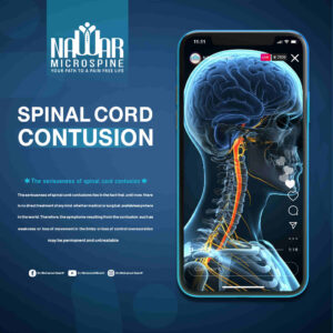
A Hidden Threat to the Spine A spinal cord contusion is one of the most serious spinal injuries, as it threatens a person’s ability to move and feel sensations. If

A Revolution in Precision Spine Surgery الرئيسية The field of spine surgery is witnessing continuous advancement, with one of the most significant breakthroughs being the use of the surgical microscope

Cervical spinal canal stenosis is one of the most common spinal problems, particularly among older adults. It causes pain, numbness, and tingling in the limbs and can lead to serious